
Figure 7. Post mortem analysis confirms the presence of mutant IDH1 and 2-HG.
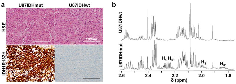
(a) U87IDHmut (left) and U87IDHwt (right) tumors had a similar histologic appearance and were densely cellular on hematoxylin and eosin staining (H&E, top row). Immunostaining for mutant IDH1 (IDH1R132H, bottom row) demonstrated robust, diffuse positivity in U87IDHmut (left) tumors and no positive staining in U87IDHwt (right) tumors (magnification ×200, scale bar 100μm). (b) 1H MR spectrum of U87IDHwt (top) and U87IDHmut (bottom) tumor lysates acquired at 14 Tesla. The multiplets characteristic of 2-HG can be seen in the U87IDHmut spectrum, not in the U87IDHwt (H3′=1.85ppm; H3=2.00ppm; H4,4′=2.25ppm; see Figure 2 for proton assignments). The level of 2-HG in U87IDHmut tumor extracts was 9.8±1.6 μmol per gram of tumor (n=3).