Figure 2.
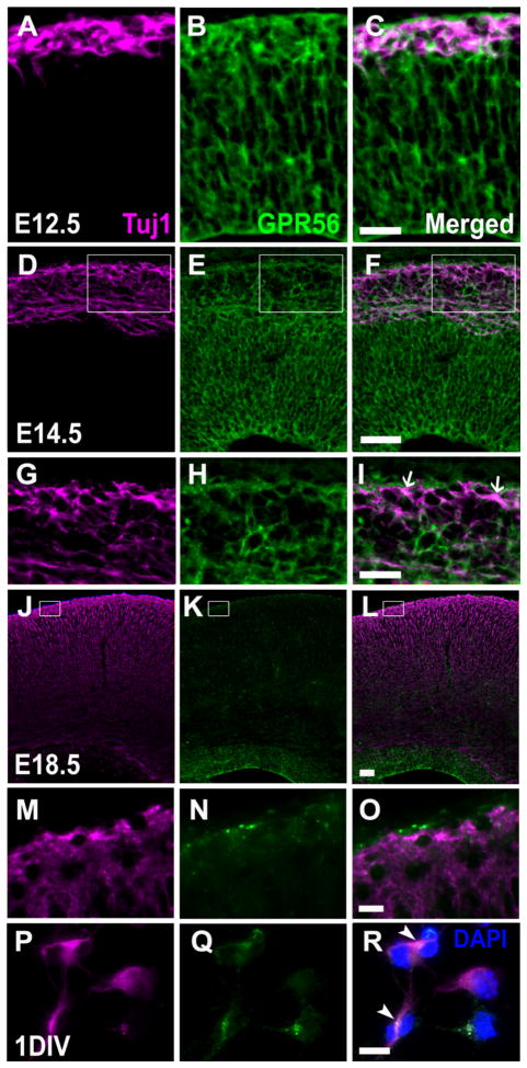
Postmitotic neurons express GPR56. A–F, J–L: Antigens were retrieved for either 8 minutes (E12.5), 12 minutes(E14.5), or 14 minutes (E18.5). Double immunostaining of Tuj1 (magenta) and GPR56 (H11, green) on coronal sections of E12.5 (A–C), E14.5 (D–F), and E18.5 (J–L) neocortex. G–I, M–O: Higher magnification of boxed regions in D–F and J–L. GPR56 is expressed in the Tuj1-positive cell population, most prominently in the preplate at E12.5. By E14.5, only scattered subpial neurons remain positive for GPR56 (arrows in I). P–R: Double immunostaining of Tuj1 and GPR56 on 1DIV progenitor cells fixed in 95% ethanol and 5% glacial acetic acid for 10 minutes at −20°C. Some Tuj1-positve neurons express GPR56 (arrowheads in R). Scale bar = 25 μm in C (applies to A–C) and I (applies to G–I); 50 μm in F (applies to D–F) and L (applies to J–L); 10 μm in O (applies to M–O) and R (applies to P–R).

