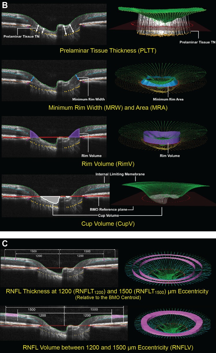
Figure 2.
(B, C) The SDOCT parameters grouped by target tissue (continued). (B) ONH neural tissues. Neural tissue parameters are designed to detect neural tissue changes that occur either due to neural tissue damage or secondary to connective tissue deformation. PLTT is measured as the normal from the tangent to the ALCS to the ILM (green line, top). The MRW (blue arrows) is measured at each delineated BMO point (red) as the minimum distance to ILM (upper middle). When viewed in a 3-D domain, MRW can be translated into minimum rim area (MRA). Rim volume (purple) is calculated from the volume bounded by ILM (green), BMO reference plane (red), and perpendicular line through the BMO (lower middle). Cup volume (grey) is generated from the volume between ILM B-spline surface and the BMO reference plane (bottom). (C) Non-standard peripapillary RNFL. RNFLT1200 is measured on either side of the posterior RNFL boundary (turquoise line) at ILM points that are 1200 μm from the centroid of the 80 delineated BMO points (the BMO centroid). Similarly, RNFLT1500 is measured at 1500 μm from the BMO centroid (top). The volume between RNFLT1200 and RNFLT1500 is defined as RNFL volume (pink, bottom).

