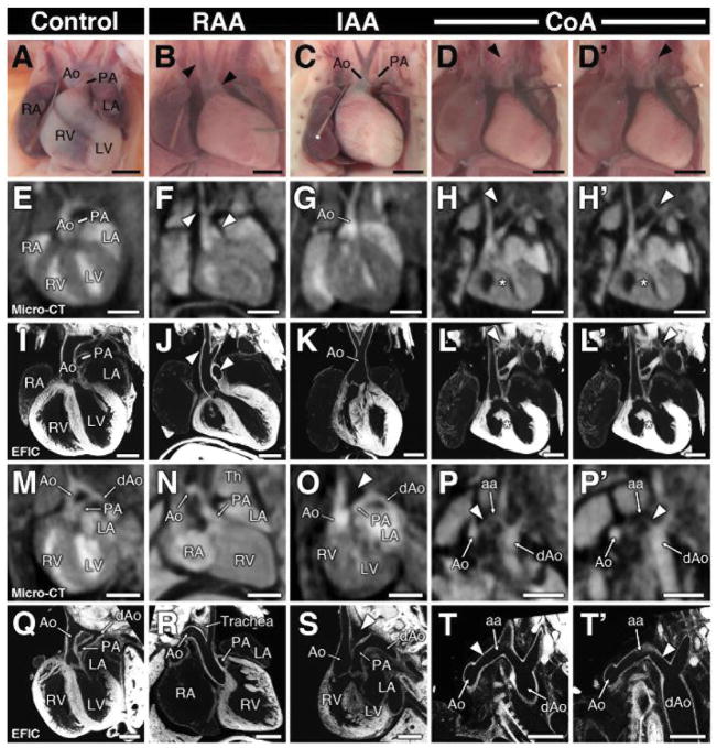
Figure 2. Aortic arch anomalies.
Visualization of aortic arch by necropsy (A–D′) and by micro-CT and EFIC imaging in the coronal (E–L′) and frontal-oblique plane (M–T′). Aortic arch anomalies detected by micro-CT include right aortic arch (RAA; B, F, J, N, R), interrupted aortic arch (IAA; C, G, K, O, S), and coarctation (CoA; D, D′, H, H′, L, L′, P, P′, T, T′). Arrowheads in panels B, F, J denote right aortic arch and orientation of the pulmonary artery. Note the aortic arch artery in panel R descends to the right of the trachea. An arrowhead in (O,S) depicts the interrupted segment of the aortic arch, with the ductus arteriosus extending and connecting to the descending aorta. Two regions of aortic coarctation in the same heart are shown in two separate panels (columns D, D′). In (P′, T′), the image orientation was tilted to show the descending aorta (P′,T′).
Scale bars = 1mm (A–H′, M–P′), and 0.5mm (I–L′, Q–T′).
aa = aortic arch; dAo = descending aorta; Th = thymus.

