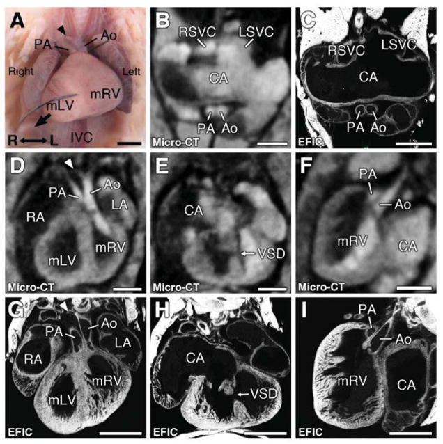
Figure 8. Complex congenital heart disease associated with heterotaxy.
Micro-CT showed heart with dextrocardia (D, G) with muscular VSD (E,H), hypoplastic right aortic arch (see arrowhead in D), and DORV with the aorta and pulmonary trunk arising from the morphologic right ventricle (D, F). This was confirmed by EFIC imaging (G,H,I). Atrial appendages showed bilateral superior vena cava (RSVC, LSVC in panel B), indicating right atrial isomerism, which EFIC histology showed was a common atrium (C).
Scale bars = 1mm.
Ao = aorta; CA = common atrium; IVC = inferior vena cava; LA = left atrium; LSVC = left superior vena cava; mLV = morphological left ventricle; mRV = morphological right ventricle; PA = pulmonary artery; RA = right atrium; RSVC = right superior vena cava; VSD = ventricular septal defect.

