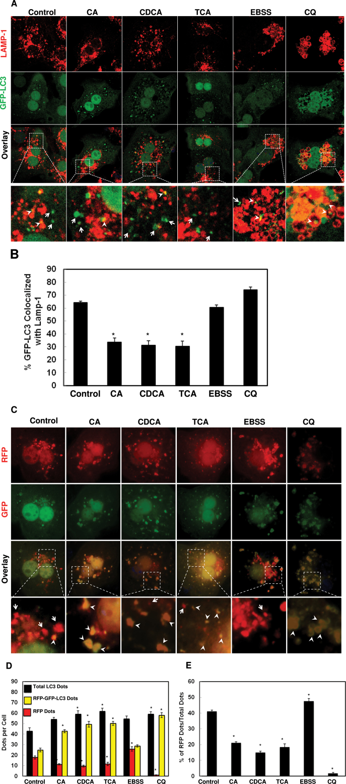
FIG. 5.
BAs impaired autophagosomal-lysosomal fusion. A, Primary hepatocytes were infected with adenoviral GFP-LC3 (100 viral particles per cell) overnight and then treated with 100 µM of BAs, or 20 µM of CQ or EBSS for 6h. Cells were immunostained for LAMP-1 followed by confocal microscopy. Representative images are shown, and the lower panels are enlarged images from the boxed areas. Arrow heads: yellow dots (colocalization of GFP-LC3 puncta with LAMP-1) and arrows: GFP-LC3 puncta only. B, Percentage of GFP-LC3 puncta colocalized with LAMP-1 (>20 cells were counted in each experiment from at least three independent experiments). C, Primary hepatocytes were transfected with RFP-GFP-LC3 plasmid for 24h and then treated as in (A). Representative images are shown, and the lower panels are enlarged images from the boxed areas. Arrow heads: yellow dots (RFP-GFP-LC3 puncta) and arrows: RFP-LC3 puncta only. D, Total RFP-LC3 and RFP-GFP-LC3 puncta were quantified and percentage of RFP-LC3 only puncta was calculated (E) (>20 cells were counted in each experiment from at least three independent experiments).

