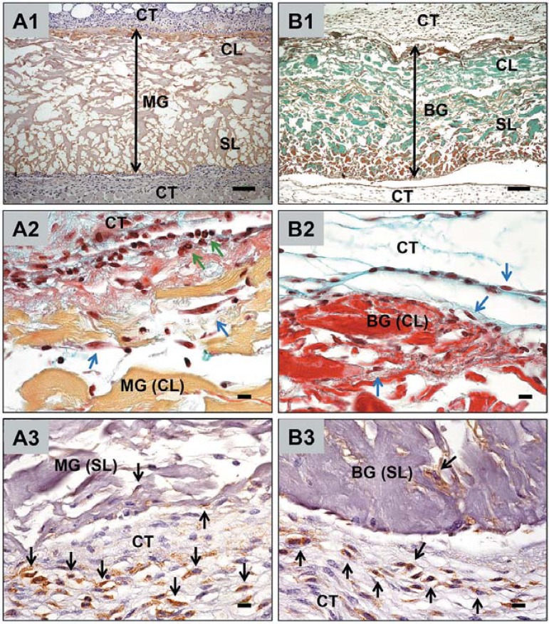
Figure 5.
Cellular interactions with the materials within the subcutaneous connective tissue (CT) of CD-1 mice. A1-3 show the tissue reactions to the Mucograft (MG) matrix, B1–3 show the tissue reaction to the BioGide (BG) membrane. Magnifications: A1 and B1 x100; (scale bar=100 μm); A2-3 and B2-3 x600; (scale bar=10 μm). CL=compact layer, SL=spongy layer. Blue arrows indicate fibroblasts (A2, B2), green arrows indicate granulocytes (A2), black arrows indicate macrophages (A3, B3)

