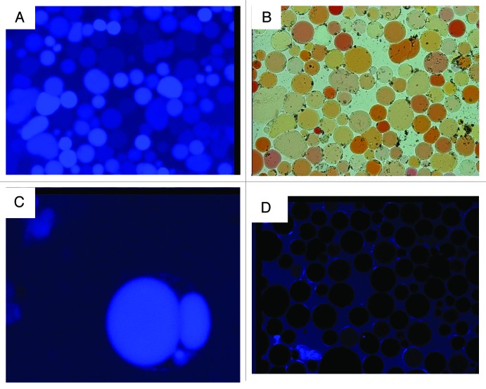
Figure 1. Red beet protoplasts isolated from tissue incubated in the presence and absence of the membrane-impermeable, endocytic-marker blue-fluorescent Alexa-350.(A) Fluorescent micrograph of protoplasts isolated from tissue incubated in 200 mM sucrose and 100 μM Alexa-350. (B) Light micrograph of (A). (C) High magnification fluorescent micrograph of a protoplast demonstrating Alexa-350 blue fluorescence in the vacuole and vacuole derived vesicles. (D) Fluorescent micrograph of protoplasts prepared from control tissue incubated in betaine.

An official website of the United States government
Here's how you know
Official websites use .gov
A
.gov website belongs to an official
government organization in the United States.
Secure .gov websites use HTTPS
A lock (
) or https:// means you've safely
connected to the .gov website. Share sensitive
information only on official, secure websites.
