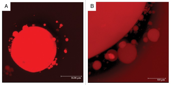
Figure 3. 3D reconstruction of Z-stack fluorescent images of red beet protoplasts isolated from tissue incubated 24 h in 200 mM sucrose. Given the wide fluorescence spectrum of betacyanin, samples were examined under an emission range of 620–650 nm. (A) Entire protoplast showing vacuole and vacuole-derived vesicles of a wide range of sizes. (B) Close up of a section of a protoplast. The dark region represents the cytosol, whereas the red structures are the vacuole, vacuole-derived vesicles and external medium still fluorescing from residual dye.

An official website of the United States government
Here's how you know
Official websites use .gov
A
.gov website belongs to an official
government organization in the United States.
Secure .gov websites use HTTPS
A lock (
) or https:// means you've safely
connected to the .gov website. Share sensitive
information only on official, secure websites.
