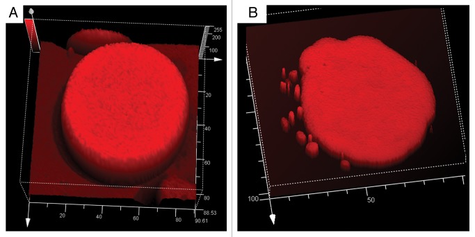
Figure 4. Topographic fluorescence analysis of red beet protoplasts isolated from tissue incubated 24 h in 200 mM sucrose at two different stages. (A) Protoplast from dormant beet hypocotyls with a large central vacuole. (B) Protoplast from tissue incubated in 200 mM sucrose for 24 h. Fluorescence intensity of vacuole-derived vesicles is similar to that emanating from the vacuole.

An official website of the United States government
Here's how you know
Official websites use .gov
A
.gov website belongs to an official
government organization in the United States.
Secure .gov websites use HTTPS
A lock (
) or https:// means you've safely
connected to the .gov website. Share sensitive
information only on official, secure websites.
