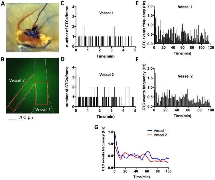
Figure 4. Imaging of circulating tumor cells in an awake, freely behaving animal using the mIVM.
(A) Photograph of the animal preparation: Following tail-vein injection of FITC-dextran for vessel labeling and subsequent injection of 1×10⁶ 4T1-GL labeled with CFSE, the animal was taken off the anesthesia and allowed to freely behave in its cage while CTCs were imaged in real-time. (B) mIVM image of the field of view containing two blood vessel, Vessel 1 of 300 µm diameter and Vessel 2 of 150 µm diameter. (C, D) Quantification of number of CTCs events during 2h-long awake imaging, using a MATLAB image processing algorithm, in Vessel 1 (C) and Vessel 2 (D). (E, F) Computing of CTC dynamics: average CTC frequency (Hz) as computed over non-overlapping 1 min windows for Vessel 1 (E) and Vessel 2 (F) and (G) Second-order smoothing (10 neighbor algorithm) of the data presented in (E, F).

