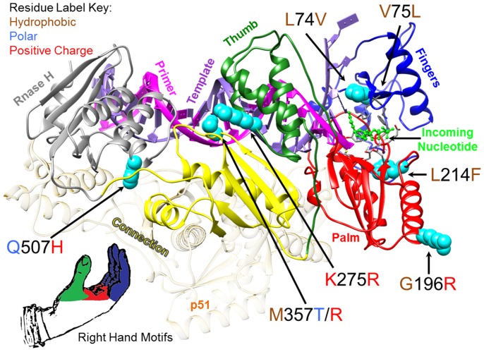
Figure 1. HIV-1 Reverse Transcriptase (RT) showing amino acid substitutions detected in RT-SHIV high virus load rhesus macaques.
HIV-1 RT p66 subunit ribbon diagram depicted with the most frequently detected amino acid substitutions as cyan-colored spheres. The domains of the RT active site are colored to correspond to the model of RT analogous to a human right hand with the fingers domain as dark blue, the palm domain as red, and the thumb domain as green. The X-ray crystal structure PDB ID: 1RTD [45] of pre-catalytic, wild type HIV-1 reverse transcriptase in complex with double stranded DNA and incoming nucleotide was used to make the image.

