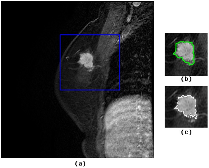
Figure 1. Segmentation of a sample breast lesion on MRI, confirmed as Invasive ductal carcinoma, for a 50 year old woman.
(a) Area including a suspicious breast lesion is highlighted by a blue rectangle; (b) Initial segmentation result on (a) by using FCM-based method; (c) Final segmented lesion after GVF snake model initialized from (b).

