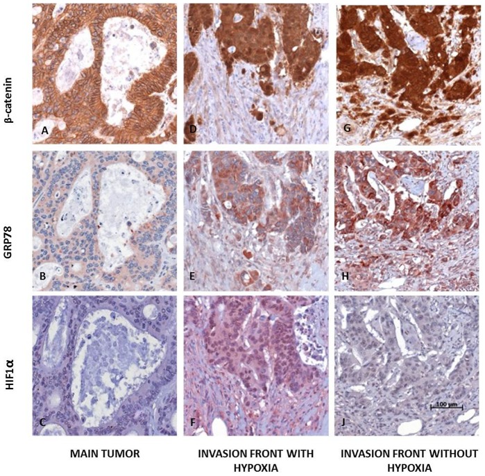
Figure 3. Human colorectal carcinomas with EMT show ER stress independent of HIF1α.
In central tumor areas of human CRCs β-catenin was typically localized at the cell membrane (A) whereas only a weak staining was observed for cytoplasmic GRP78 (B) and HIF1α staining was found to be negative (C). At the invasion front strong nuclear β-catenin was detectable indicating EMT (D, G). In corresponding regions strong cytoplasmic GRP78 expression was found (E, H). In some of the cases an intense nuclear HIF1α staining was observed (F, with hypoxia), but not in others (I, without hypoxia) (magnification 200×; scale bar: 100 µm).

