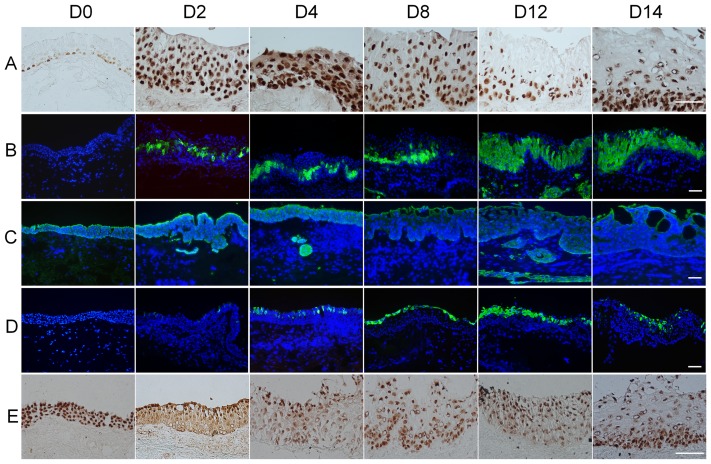
Figure 2. Conjunctival epithelial squamous metaplasia in airlift cultures.
(A) P63 staining showed positive nuclei in all basal cells and some suprabasal cells in normal human conjunctiva (D0). In the airlift culture, p63 expression increased throughout the conjunctival epithelial layers at D2, while the superficial cell layers lost p63 expression from D12 to D14. (B) K16 positive cells were not present in normal conjunctival epithelium (D0). In the airlift culture, K16 positive cells emerged in the suprabasal layer from D2 and gradually increased and spread to superficial layers from day 4 to day 14. (C) K19 expression in the full thickness freshly isolated ex vivo conjunctival epithelial cells (D0). K19 expression gradually decreased in the airlift culture after 4 days of cultivation. There were K19 positive cell clusters in the conjunctival stroma. (D) K10 staining was negative in normal conjunctiva before culturing (D0), but became positive in superficial cell layers at day 2 and day 4, and gradually increased to lower layers from day 8 to day 14. (E) Pax6 expressed in full thickness of normal conjunctival epithelial cells (D0), and it decreased in the basal and supra-basal layers in airlift culture from D2 to D14. Bars represent 100 µm.

