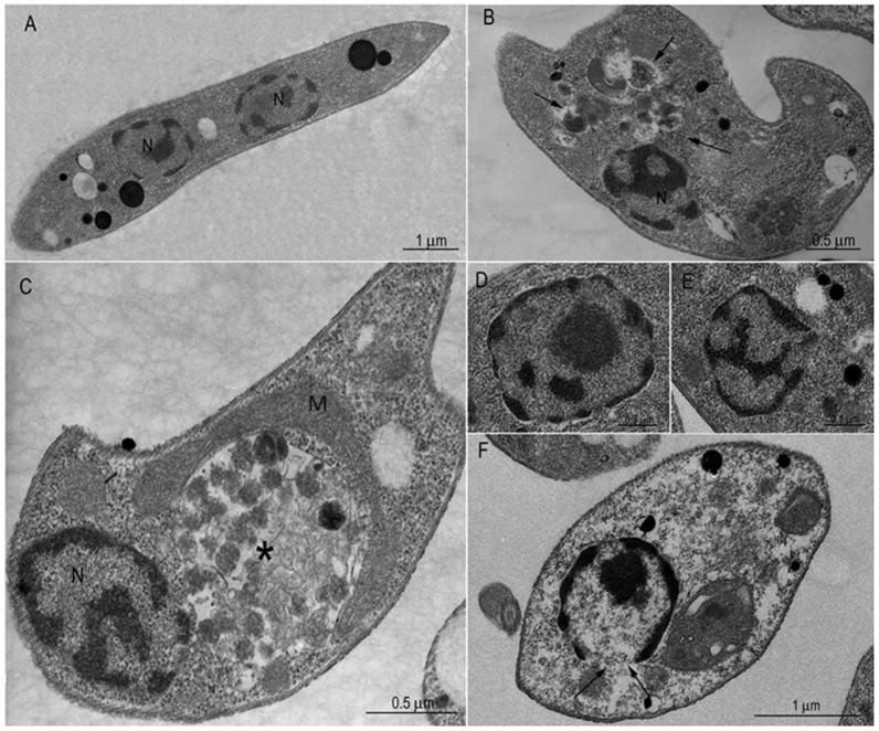
Figure 3. Transmission electron microscopy of Leishmania amazonensis promastigotes after treatment with MDL28170.
(A) Control, untreated cell: note the elongated body and the normal aspect of the intracellular organelles (N, nucleus). In MDL28170-treated promastigotes both at IC50 (B–C) and two times the IC50 (2×IC50) doses (D–E), the ultrastructural changes show intense vacuolization in the cytoplasm (arrows in B), disorganization of compartments of the endocytic pathway containing multivesicular bodies-like vesicles (C, asterisk), altered chromatin condensation pattern (D–F) and apparent loss of nuclear integrity (F, small arrows). M, mitochondrion.

