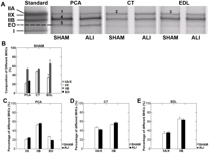
Figure 7. MyHC isoforms in the limb and laryngeal muscles of WT mice.
A. Silver stained gels of separated MyHC isoforms of representative SHAM and ALI mice in the PCA, CT and EDL muscles. The PCA muscle lysate included an additional band (band 5), which was not present in the standard, CT or EDL lysates. Mass spectroscopy analysis (see table 1) revealed band 5 to be MyHC-Extraocular (EO) isoform. Distribution of MyHC isoforms (B) in each muscle was quantified by the percentage of integrated density of each bands within each muscle (n = 5/group). No evidence of MyHC isoform switching was present in ALI mice at day 3 in the PCA (C), CT (D) or EDL (E) muscle.

