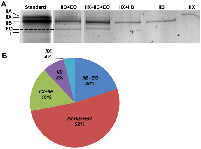
Figure 8. Identification of muscle fiber types in the PCA by MyHC isotyping of single fibers.
80 single fibers from the PCA were dissociated and run in individual lanes and silver stained for identification of MyHC isoforms present within each muscle fiber. A. Representative examples of the differing MyHC isoforms present within single fibers of the PCA muscle. B. Quantification of the percentage of MyHC fiber types present in the PCA muscle revealed 88% of fibers co-expressed multiple MyHC isoforms and 72% of muscle fibers contained MyHC-EO.

