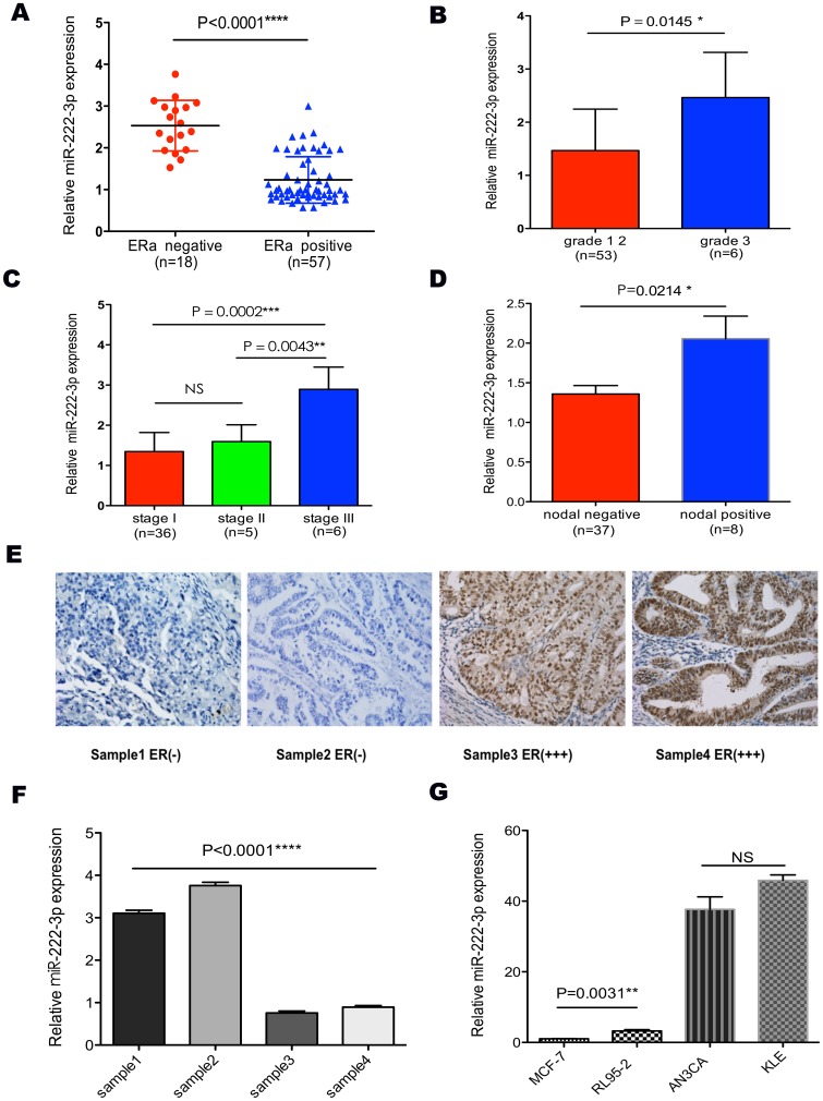
Figure 1. The expression of miR-222-3p correlated with ERα and clinicopathological parameters in endometrial carcinoma.
(A) MiR-222-3p expression level was much higher in ERα-negative ECs than in ERα-positive samples. (B, C and D) Expression of miR-222-3p across different grades, stages and nodal metastasis status. MiR-222-3p expression level was positively correlated with poor clinicopathological parameters. (E and F) MiR-222-3p and ERα expression were inversely related in ECs and normal endometrium tissues. These tissues were analyzed for miR-222-3p expression by TaqMan based qRT-PCR, followed by immunohistochemistry for ERα as described in “Materials and Methods”. (G) Expression of miR-222-3p in cells with different ERα status. MCF-7 and RL95-2 cells were ERα-positive, while KLE and AN3CA cells were ERα-negative. Conversely, miR-222-3p expression was negative-related with ERα status. Bars are standard deviation (SD). The experiments were repeated three times. ** P<0.01 vs. RL95-2, MCF-7. ****P<0.0001 vs. RL95-2, KLE, AN3CA. * P<0.05, ** P<0.01, *** P<0.001, **** P<0.0001.

