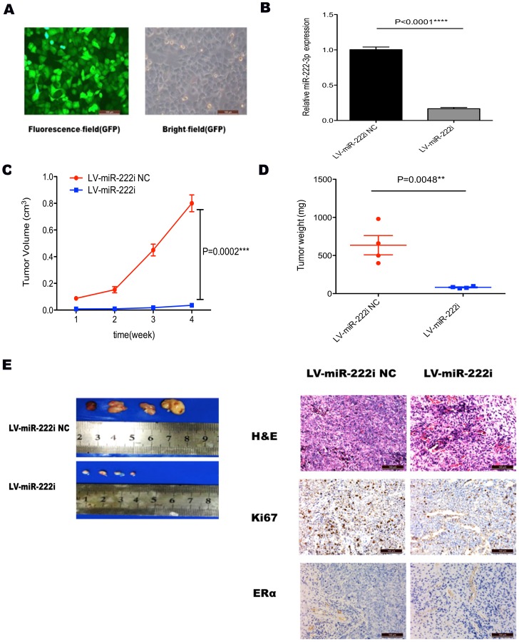
Figure 5. Tumorigenicity assay in nude mice.
(A) Stable transfection of AN3CA cells with LV-miR-222i and LV-miR-222i NC. The percentage of transfected cells with fluorescence was >95%. (B) qRT-PCR analysis of miR-222-3p expression after transfecting with LV-miR-222i and LV-miR-222i NC in AN3CA cells. **** P<0.0001. (C) Tumor growth in nude mice. AN3CA cells with different treatment were injected subcutaneously into the interscapular area of nude mice. The short and long diameters of the tumors were measured weekly and tumor volumes (cm3) were calculated. *** P<0.001. (D) Weight of tumors in nude mice. **P<0.01 vs. either no transfection or control vector. (E) The nude mice with tumor formation and representative HE staining histopathologic image of tumor tissues in mice (upper panel, 200×). The expression of Ki67 and ERα in the tumor was detected by immunohistochemical techniques (200×).

