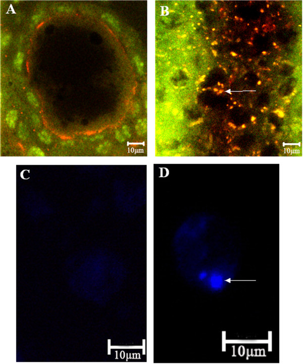
Figure 3.

Confocal and fluorescent micrographs of control (A, C) and treated (B, D) terminal follicles of T. molitor. In control follicular cells stained with orange acridine, lysosomes are not detected and nuclei of follicular cells (green colour) and oocyte (dark colour) are visible (A). In the follicular cells of treated females orange acridine stain shows many lysosomes (yellow colour, arrow) (B). In isolated control follicular cells stained with MDC, autophagic vacuoles are not detected (C). In isolated follicular cell of treated females stained with MDC arrow shows autophagosome (blue colour) (D). Females were injected with saline (control) or 10 nmole Neb-colloostatin (treated) on day 3 and assayed on day 4.
