
FIGURE 1.
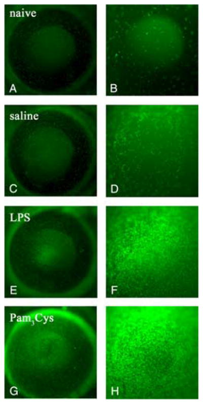
Cellular infiltration in TLR4−/− mice corneas reconstituted with eGFP+ C57BL/6 bone marrow cells. TLR4−/ − mice were irradiated and reconstituted with bone marrow from eGFP+ C57BL/6 mice. After 4 wk, corneas were abraded and exposed to saline, LPS, or Pam3Cys. After 24 h, in vivo epifluorescence microscopy images were captured at magnification ×32 (A, C, E, and G) or ×80 (B, D, F, and H). Representative images are shown from naive (A and B), saline treated (C and D), LPS-treated (E and F ), and Pam3Cys treated corneas (G and H). eGFP+ cells were present in the host corneas of naive mice (A, total cornea; B, central cornea) and a mild infiltrate of eGFP+ cells was observed in saline treated control mice, trauma induced response (C, whole cornea; D, central cornea). In LPS treated corneas, more eGFP+ cells were visible in both the limbal region (E) and in the central cornea (F ) compared with saline treated corneas. Pam3Cys treated corneas induced recruitment of eGFP+ cells in the limbal region (G) and in the central cornea (H). These experiments were repeated three times with four mice per group.