
FIGURE 3.
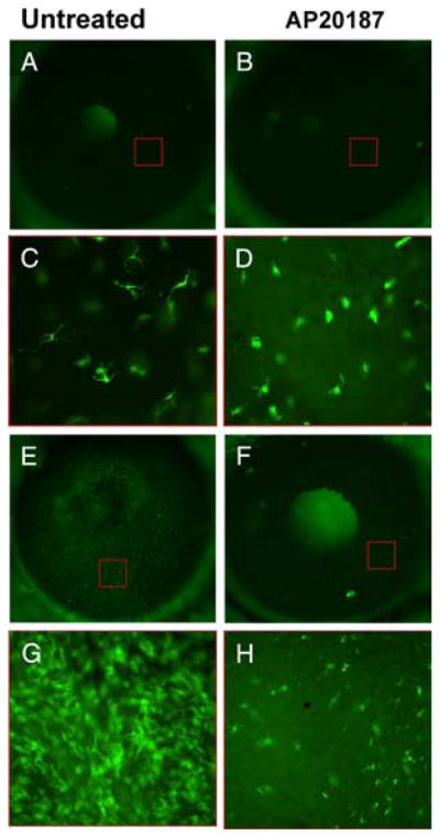
Depletion of c-fms– expressing eGFP+ cells (macrophage and DCs) in Mafia mice. Mafia mice were left untreated (A, C, E, and G) or were injected i.p. with the AP20187 dimerizer to deplete c-fms cells (B, D, F, and H). Corneas were viewed either by epifluorescence of the whole eye (A, B, E, and F), or at higher magnification after dissection of the cornea (C, D, G, and H). Before corneal stimulation (A–D) resident eGFP+ cells were detected in the corneas of untreated mice (A and C); however, there was an almost complete loss of typical dendriform intraepithelial Langerhans like cells in mice treated with the dimerizer (B and D), although eGFP+ cells were present in the corneal stroma. Corneas were abraded and stimulated with LPS as before, and examined after 24 h (E–H). In Mafia mice not injected with the dimerizer, there was an intense infiltrate of eGFP+ cells in all regions of the cornea (E and G), whereas there was an obvious reduction in the numbers of eGFP+ cells in dimerizer-treated mice (F and H). At the higher magnification, cells are evident in the corneal epithelium and stroma (G and H). Red boxes in A, B, E, and F indicate approximate regions where the higher magnification images were obtained. Images are representative of three repeat experiments with five mice per group. Original magnification A, B, E, and F, ×20; C and D, ×400. (The central bright area in panels A, B, and F is the mouse lens seen through the pupil.)