
FIGURE 5.
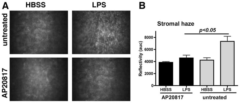
The effect of depletion of c-fms-expressing cells on corneal thickness and haze as visualized and measured by in vivo confocal microscopy. A, Representative images of cellular infiltrate in untreated Mafia mice compared with AP20187 treated Mafia mice. B, Quantitative differences in stromal haze (reflectivity). Similar results were found in three repeat experiments with five mice per group.