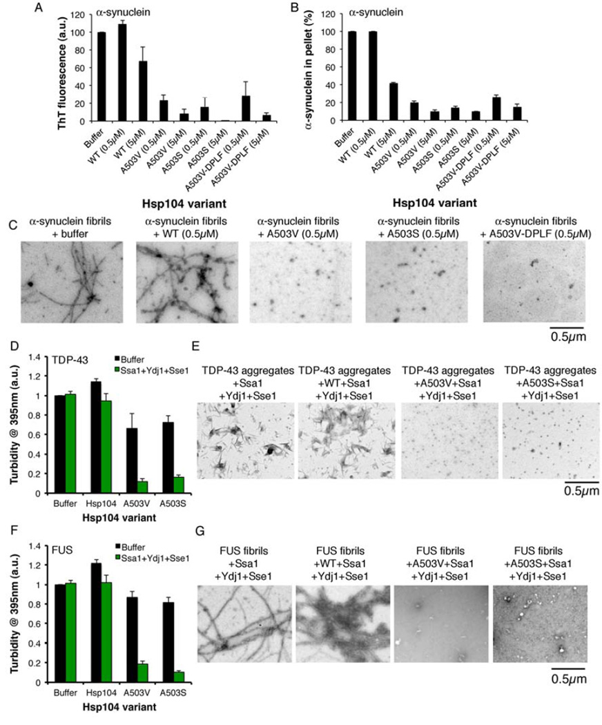
Figure 7. Potentiated Hsp104 variants disaggregate preformed α-syn, TDP-43, and FUS fibrils more efficaciously than Hsp104WT.
(A–C) α-syn fibrils were incubated without or with Hsp104WT, Hsp104A503V, Hsp104A503S, or Hsp104A503V-DPLF for 1h at 30°C. Fiber disassembly was assessed by ThT fluorescence (A), sedimentation analysis (B), or (C) EM (Bar, 0.5µm). (A, B) Values represent means±SEM (n=2). (D, E) TDP-43 aggregates were incubated with buffer, Hsp104WT, Hsp104A503V, or Hsp104A503S plus or minus Ssa1, Ydj1, and Sse1 for 1h at 30°C. (D) Aggregate dissolution assessed by turbidity. Values represent means±SEM (n=3). (E) Aggregate dissolution assessed by EM. Bar, 0.5µm. (F, G) FUS aggregates were incubated with buffer, Hsp104WT, Hsp104A503V, or Hsp104A503S plus or minus Ssa1, Ydj1, and Sse1 for 1h at 30°C. (F) Aggregate dissolution assessed by turbidity (absorbance at 395nm). Values represent means±SEM (n=3). (G) Aggregate dissolution assessed by EM. Bar, 0.5µm.

