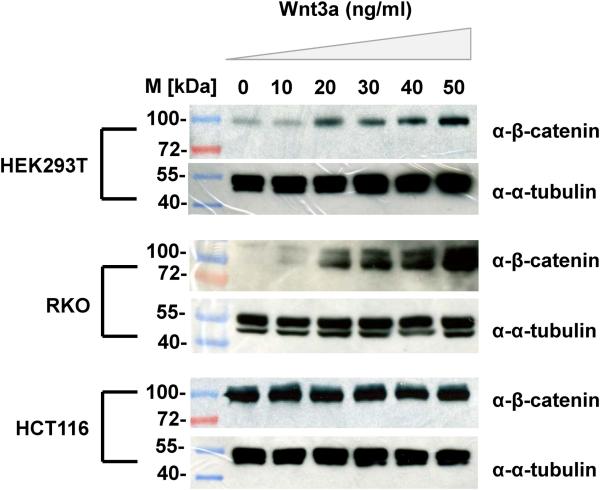
Figure 2.
Calibrating the Wnt-response in different cell lines (HEK293T, RKO, HCT116). Immunoblots show β-catenin protein expression. Cells were treated with varying doses of Wnt3A (0-50ng/ml), incubated for 24 hours and twenty micrograms of total proteins extracted from each cell lysate were subjected to SDS-PAGE followed by immunoblotting with β-catenin antisera or α-tubulin (as loading control). M refers to standard protein marker.

