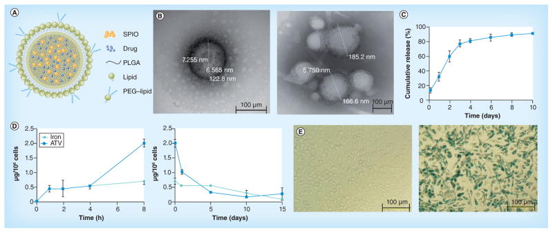
Figure 1. Development of small magnetite antiretroviral therapy nanoparticles.

(A) The schematic structure of lipid-coated PLGA small magnetite antiretroviral therapy (SMART). ATV and SPIO are well distributed within the PLGA matrix to form the core of SMART. The PLGA core is coated with lipid monolayer to form the shell of SMART. (B) Representative transmission electron micrograph of a single SMART particle. (C) Cumulative release of ATV in isotonic solution. (D) Time course of uptake (upper panel) and retention (lower panel) of SMART in monocyte-derived macrophages (MDMs). MDMs were treated with 100 μM SMART (based on ATV content) for 1-, 2-, 4- and 8-h uptake studies. For cell retention, MDMs were treated with 100 μM SMART for 8 h, cell culture media was changed and cells were cultured for an additional 15 days. The cell lysates at indicated times were analyzed by HPLC and inductively coupled plasma mass spectrometry for ATV and magnetite quantification, respectively. Data represent the mean ± standard error of the mean (n = 3), for each time point. (E) Prussian blue staining of MDMs. MDMs were treated with phosphate-buffered saline (negative control, upper panel) or 100 μM SMART (lower panel) for 24 h, and then fixed with 2% formalin/2.5% glutaraldehyde and stained with 5% potassium ferrocyanide/5% hydrochloric acid (1:1).
ATV: Atazanavir; SPIO: Superparamagnetic iron oxide; PLGA: Polylactic-co-glycolic acid.
