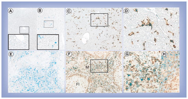
Figure 6. Immunohistology of Iba-1 staining and Prussian blue staining in liver and spleen.

(A) Liver from control mice with Prussian blue (magnification: 200×; inset 400×). (B) Liver from small magnetite antiretroviral therapy (SMART)-treated mice with Prussian blue (magnification: 200×; inset 400×). (C) Liver from SMART-treated mice with Prussian blue and Iba-1 (magnification: 200×). (D) Enlargement from indicated section in (C). (E) Spleen from SMART-treated mice with Prussian blue (magnification: 200×). (F) Spleen from SMART-treated mice with Prussian blue and Iba-1 (magnification: 200×). (G) Enlargement from indicated section in (F). (H) Spleen from control mice with Prussian blue and Iba-1 (magnification: 400×). Livers and spleens were fixed with 10% formalin, paraffin embedded and sectioned for immunohistological analysis after the final MRI scan. Macrophages were identified by Iba1 stains (brown) and magnetite identified by Prussian blue.
Fl: Lymphoid follicle; M: Marginal zone.
