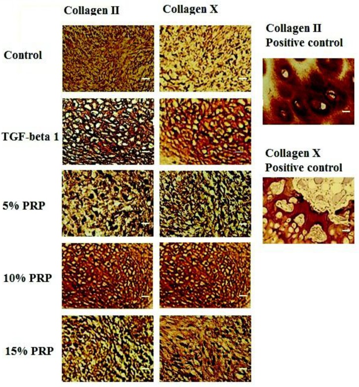
Figure 3.
Immunohistochemistry staining. First column shows the staining for type II collagen in control, TGF-β1, 5% PRP, 10 % PRP and 15 % PRP groups. Second column represents immunohistochemistry staining of different groups’ samples for type X collagen. Third column: positive control samples. Upper insert shows the human articular cartilage as a control for type II collagen and lower insert represents the human osteochondral plug as control for type X collagen. Scale bar: 20 µm

