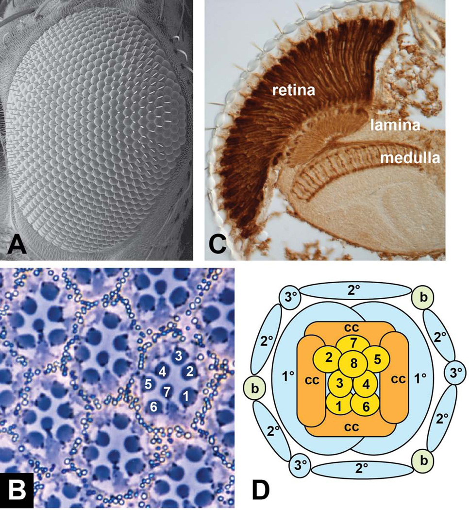
Figure 1. Structure of the adult Drosophila eye.
(A) shows a scanning electron micrograph of the surface of the eye, demonstrating the hexagonal packing of the ommatidia. (B) shows a tangential section through the eye, illustrating the characteristic trapezoidal arrangement of the rhabdomeres of photoreceptors R1-R7. The rhabdomere of R8 lies below that of R7. (C) shows a coronal section through the adult head of a fly expressing lacZ in all photoreceptors, stained with anti-β-galactosidase. This section shows the elongated shape of the photoreceptor cells in the retina and their axons extending to the lamina and medulla. (D) is a diagram of the arrangement of cell types found in each ommatidium. 1–8, photoreceptors R1-R8; cc, cone cells; 1°, 2°, 3°, pigment cells; b, mechanosensory bristle.

