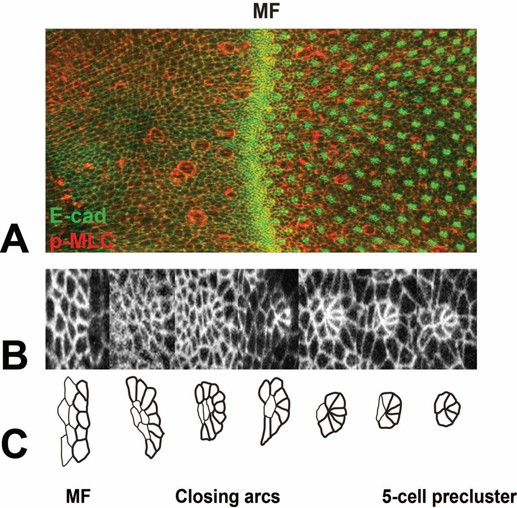
Figure 2. Pattern formation in the developing eye disc.
(A) shows part of an eye disc labeled with E-cadherin-GFP (E-cad-GFP, green) and anti-phosphorylated myosin light chain (p-MLC, red). Anterior is to the left. The morphogenetic furrow (MF) is indicated. (B) shows a series of E-cad-GFP-labeled cell clusters increasing in age from left to right, and (C) shows tracings of the same clusters. Unpatterned cells in the morphogenetic furrow transform into arcs, which close by removal of the central cells and ultimately become 5-cell preclusters. Figure kindly provided by Franck Pichaud.

