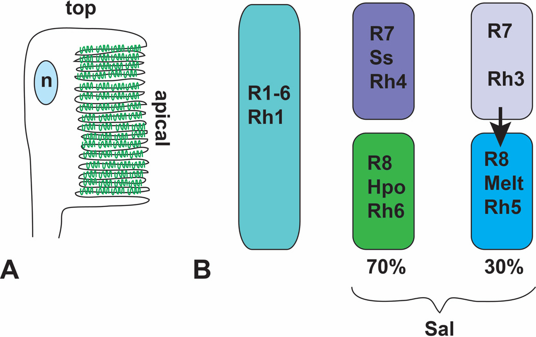
Figure 6. Terminal differentiation involves Rhodopsin expression and localization.
(A) is a diagram of the adult rhabdomere, indicating the rotation and folding of the apical surface. Rhodopsin molecules are represented in green. n, nucleus. (B) shows the distribution of the five different rhodopsins between the eight photoreceptors. All R1–6 cells express Rh1. Approximately 70% of R7 cells express Ss and Rh4. The R8 cells in the same ommatidia activate the Hpo pathway and express Rh6. In the absence of Ss, R7 cells express Rh3 and signal to the R8 cells in their ommatidia to express Melt and Rh5

