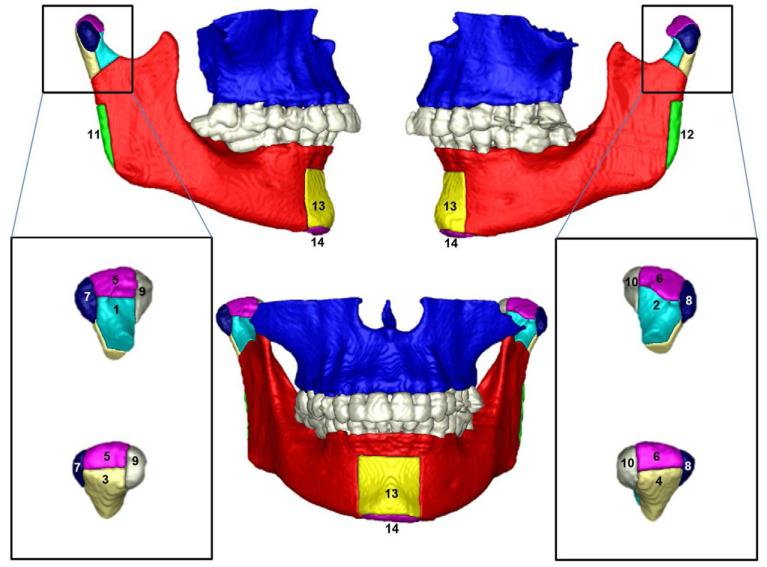
Figure 1.
Anatomic regions of interest: 1, right condyle anterior surface; 2, left condyle anterior surface; 3, right condyle posterior surface; 4, left condyle posterior surface; 5, right condyle superior surface; 6, left condyle superior surface; 7, right condyle lateral pole; 8, left condyle lateral pole; 9, right condyle medial pole; 10, left condyle medial pole; 11, right posterior border ramus; 12, left posterior border ramus; 13, anterior surface of the chin; and 14, inferior border of the mandible.

