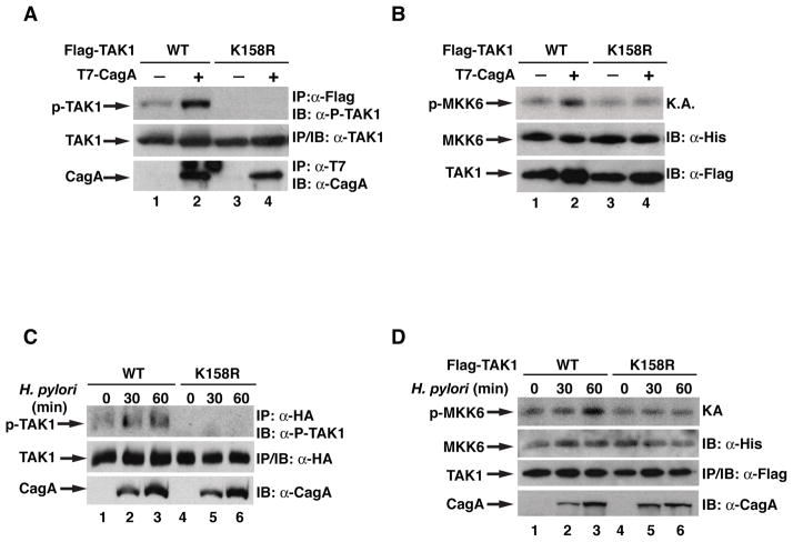
Figure 2. Ubiquitination at lysine 158 of TAK1 is important for its kinase activity.
(A) WT or K158R TAK1 were expressed in HEK293T cells with or without CagA. Flag-TAK1 immunoprecipitates were immunoblotted for phosphorylated TAK1. (B) WT or K158R TAK1 were expressed in HEK293T cells with or without CagA. Flag-TAK1 immunoprecipitates were subjected to an in vitro kinase assay using His-MKK6(K82A) as a substrate. Levels of phosphorylated MKK6, TAK1, and MKK6 are shown. (C) AGS cells expressing WT or K158R TAK1 were infected with H. pylori for indicated times. Flag-TAK1 immunoprecipitates were immunoblotted for phosphorylated TAK1. (D) AGS cells expressing WT or K158R TAK1 were infected with H. pylori for indicated times. Flag-TAK1 immunoprecipitates were subjected to an in vitro kinase assay as in (B).

