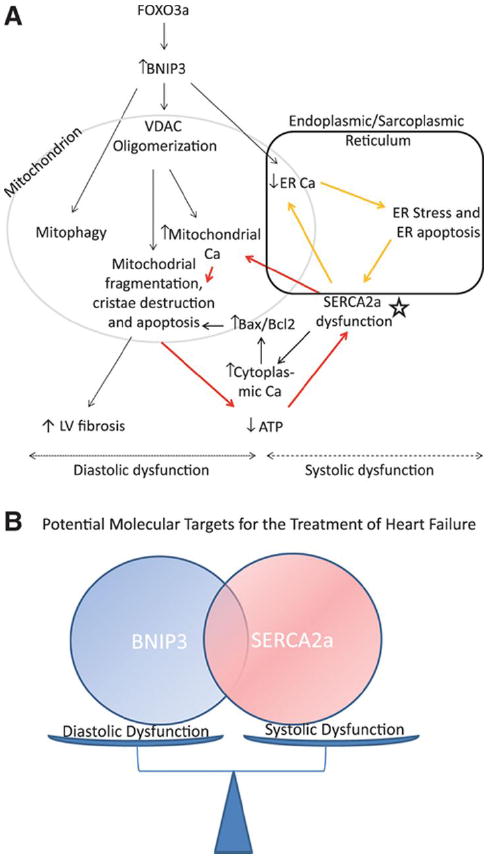
Figure 7. Schematic illustration of the mechanism by which BNIP3 induces mitochondrial damage and cell death.

7-A: The increase in BNIP3 expression leads to the oligomerization of the VDAC channels causing the shift in ER Ca2+ towards the mitochondria. This shift of ER Ca2+ has two sequelae, the first being the increase in mitochondrial Ca2+ and the second is the decrease in ER Ca2+ content. The increase in mitochondrial Ca2+ leads to mitochondrial fragmentation, mitophagy and apoptosis, decline in cardiac energetics and LV interstitial fibrosis (diastolic dysfunction). Whereas, the decrease in ER Ca2+ leads to ER stress and ER stress induced apoptosis (systolic dysfunction). In systolic HF, SERCA2a downregulation (star), which is an independent process from the increase in BNIP3 expression, contributes to the further decline in ER Ca2+ and to the further increase in mitochondrial Ca2+ leading to two vicious cycles and hence becomes the core of these two cycles as shown in the figure above (red and yellow arrows). 7-B: Cartoon highlighting the interplay between BNIP3 and SERCA2a in modulating diastolic and systolic function of the cardiomyocyte, respectively.
