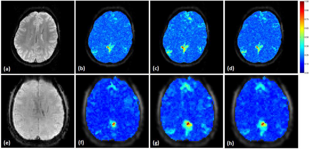
Figure 9.
Mutual information (MI) between a seed in the PCC and the original and denoised fMRI data calculated from two sets of resting state data. The MI values were normalized to the maximum MI computed from the original and denoised fMRI data in each data set, and overlaid on the corresponding slice that covers the seed region, (a) shows an individual slice from one data set where the seed was selected, (b) is the MI map calculated using the original data, (c) is the MI map computed using the data denoised by the proposed method, and (d) was from the data processed by the adaptive filtering method, (e) is an individual slice from the other data set where the seed was selected in PCC. (f)–(h) are the MI maps following the same order as (b)–(d) calculated using the original and denoised slice in this data set.

