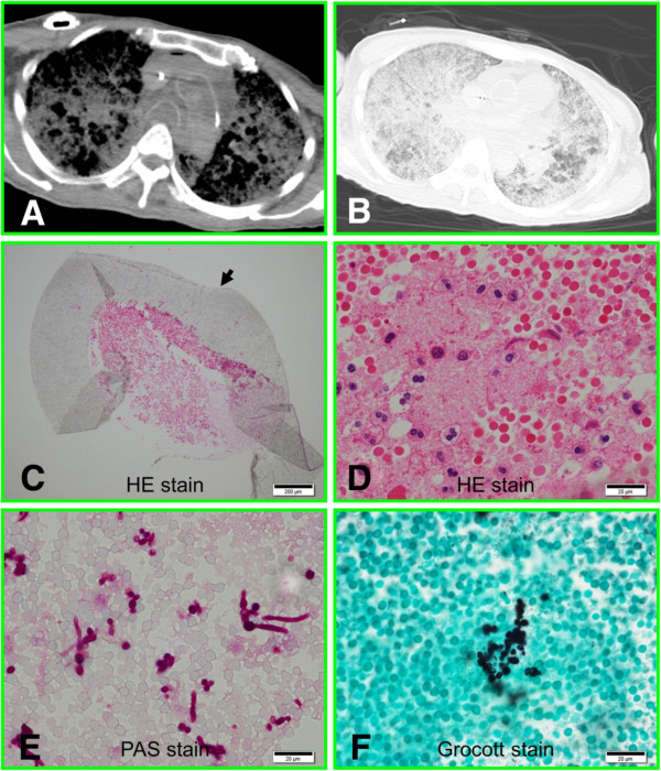
Figure 1.
Postmortem analysis. A) Postmortem CT of mediastinal window. B) Postmortem CT of lung window. C) Fibrin thrombus in the CV port tube (arrow). D) High-magnification view of the thrombus containing neutrophils and macrophages. E)Candida in the thrombus stained by the periodic acid-Schiff reagent. F) Detection of Candida by Grocott staining.

