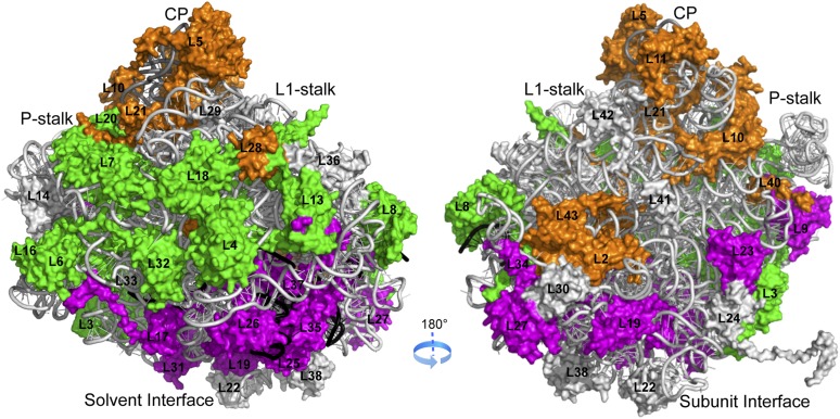
Figure 1.
Correlation between function and localization of RPLs. The location of early-acting (green), intermediate-acting (magenta), and late-acting (orange) RPLs is mapped onto the crystal structure of the yeast 60S subunit (Protein Data Bank [PDB] accession nos. 3U5D and 3U5E). rRNAs and RPLs are shown in cartoon and surface representations, respectively. 5.8S, 25S, and 5S rRNAs are colored black, light gray, and dark gray, respectively.

