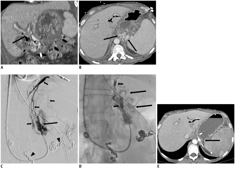
Fig. 2.
51-year-old women with necrotizing pancreatitis, portal vein thrombosis, and gastric varices.
A, B. Coronal reformatted (A) and axial (B) contrast-enhanced computed tomography (CT) scan 3 days prior to balloon occlusion retrograde transvenous obliteration (BRTO) procedure shows abrupt cut off of both right and left portal veins (arrow), representing portal vein thrombus, splenic vein thrombosis, and enhancing dilated veins (arrows) in region of gastric fundus representing gastric varices. C. Balloon occluded retrograde venogram shows filling of gastric varices (arrow) and multiple collateral veins including inferior phrenic vein (small arrows). Note previous coil embolized splenic and gastroduodenal arteries (arrowheads). Two collateral veins including inferior phrenic vein were embolized with multiple microcoils. Sclerosant was administered with occlusion balloon inflated and filling of varices. D. Spot image of gastric varices post embolization shows gastric varices with lipiodol uptake (arrows) and multiple coils (small arrows) at embolized inferior phrenic vein and small draining vein. E. Axial contrast-enhanced CT scan 6 months after BRTO procedure shows complete obliteration of gastric fundus with small residual lipiodol uptake (arrow).

