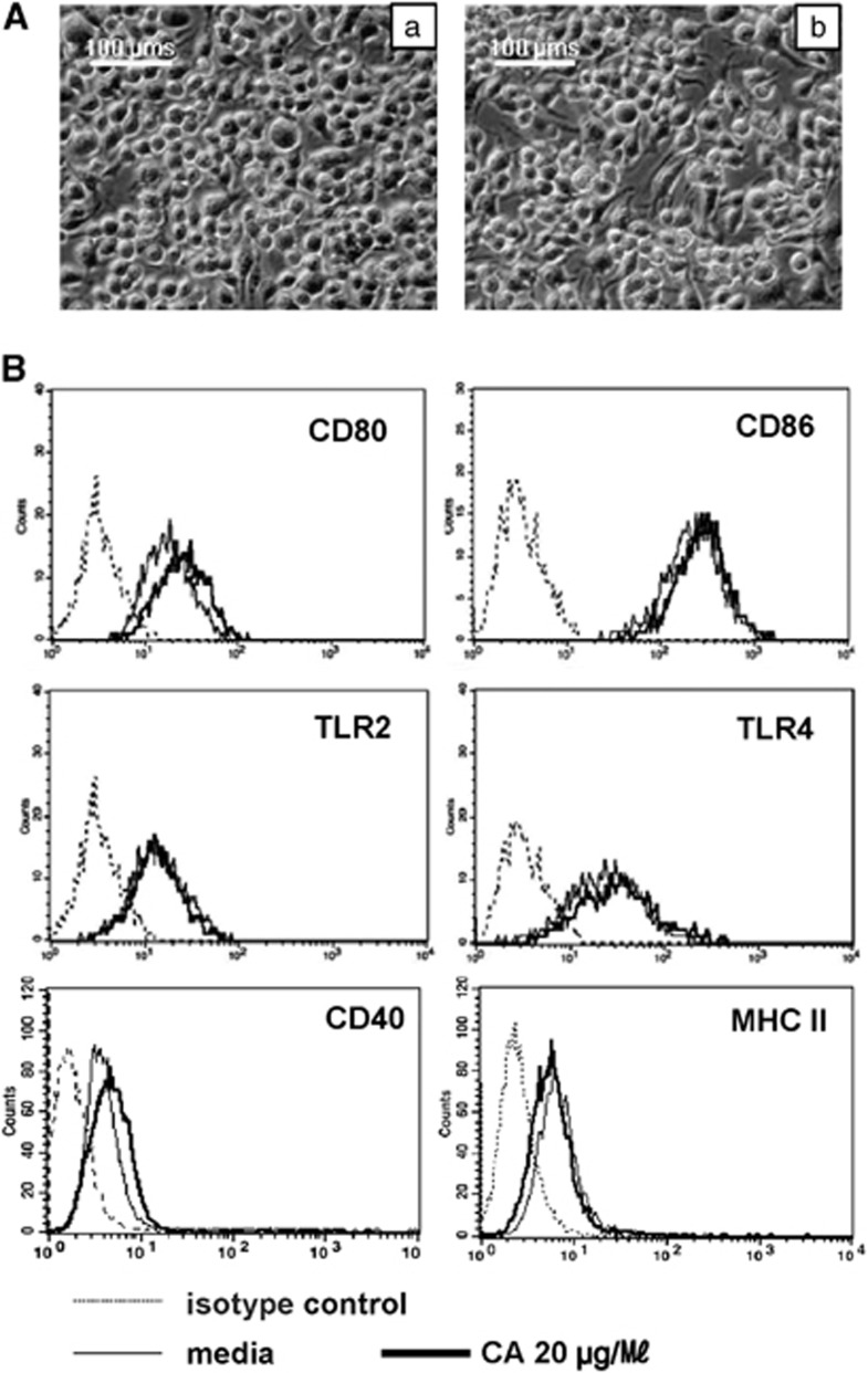
Figure 1.
Panel A shows Morphological changes of DC2.4 cells treated with (a) medium, (b) 20 μg ml−1 of crude antigen (CA). Cells were plated in six-well plates (2 × 10⁵ cells per well). After incubation for 24 h, changes in cellular morphology in each group were determined using microscopy. Images were acquired using a digital camera at × 200 magnification. This figure shows representative features of four independent experiments. Panel B shows no differences in levels of cell surface markers (CD80, CD86, Toll-like receptor (TLR)2 and TLR4) after treatment with C. sinensis crude antigen.

