Figure 4.
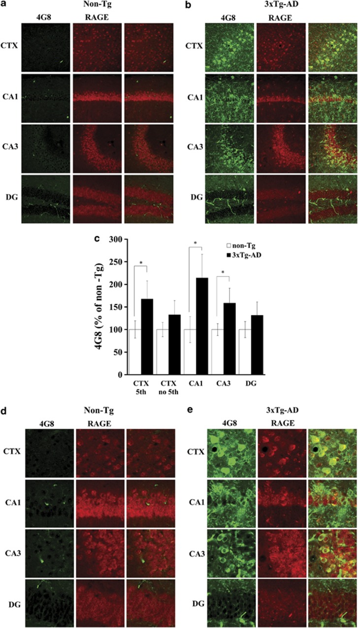
Distribution of 4G8-positive APP/Aβ and its co-localization with RAGE in aged 3xTg-AD mice. Confocal images of double-labeling with RAGE and 4G8 in the hippocampus and cortex (CTX) in aged non-Tg control (a, d) and 3xTg-AD (b, e) mouse. Immunoreactive labeling for 4G8, a monoclonal antibody detecting APP/Aβ, was increased in aged 3xTg-AD mice compared with aged non-Tg controls (c). Notably, in the cortex, the immunoreactivity of 4G8 was significantly increased only in layer 5, whereas there were no differences in other layers (c). In the hippocampus, 4G8 immunoreactivity was significantly increased in area CA1 and CA3 but not in the DG (c). Labeling for intracellular 4G8 in neurons was co-localized with RAGE expression, whereas that for extracellular 4G8 was not (e). Data are means±s.e.m. (adult: n=6; aged: n=10). *P<0.05. Scale bar=100 μm.

