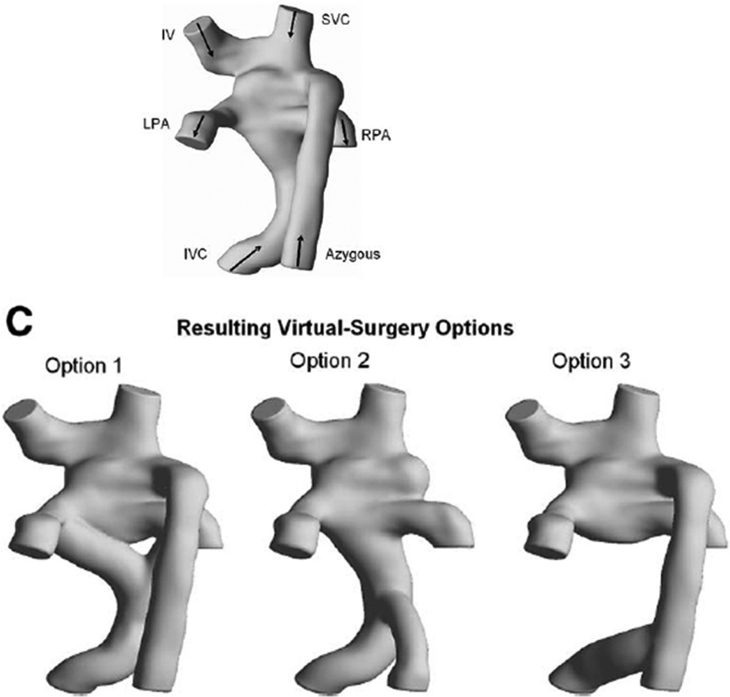
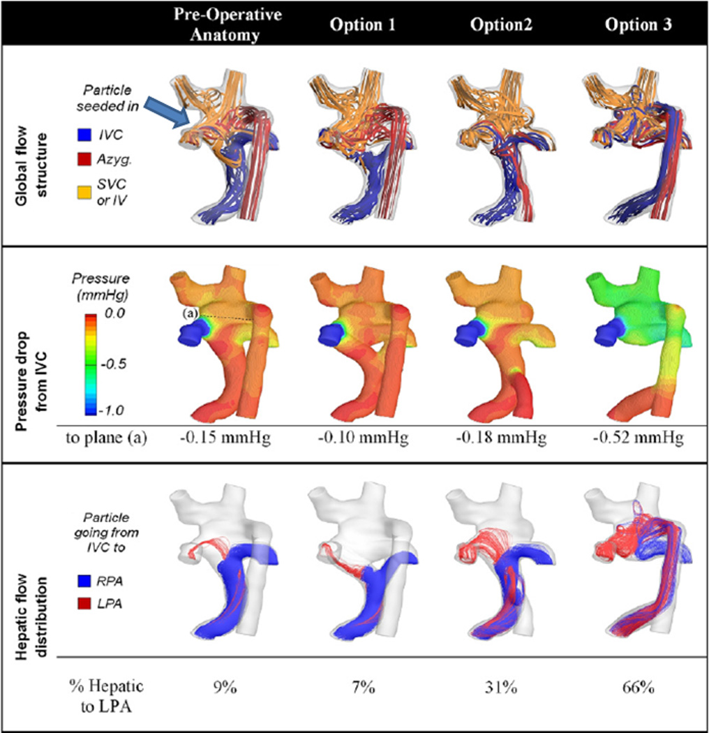
Figure 3.


Surgical planning in a Fontan patient with heterotaxy. (A) All 3D images are viewed from posterior. Original anatomy is at the top and the 3 virtual surgical options are at the bottom below the letter "C." Y graft, azygous connection to the hepatic baffle and hepatic baffle connection to the azygous are shown from left to right respectively. (B) Flow structures (top row), pressure drop (middle row) and hepatic flow (bottom row) for the original anatomy (left column) and the 3 surgical options are shown. Arrow points to the vortex formation in the original anatomy. Azyg=azygous, IV=innominate vein, IVC=inferior vena cava, LPA=left pulmonary artery, mm Hg= millimeters of mercury, RPA=right pulmonary artery, SVC=superior vena cava. From Sundareswaran KS, de Zelicourt D, Sharma S, Kanter KR, Spray TL, Rossignac J, Sotiropoulos F, Fogel MA, Yoganathan AP. Correction of pulmonary arteriovenous malformation using image-based surgical planning. J Am Coll Cardiol Img. 2009;2:1024–1030. With permission.
