
Figure 2.
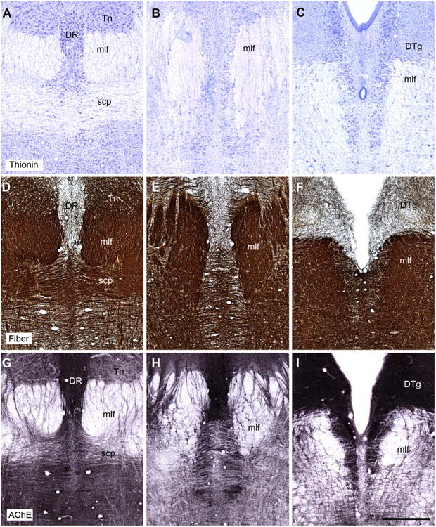
A–I: Coronal sections showing the appearance of the tegmental and raphe regions in the rhesus monkey (Macaca mulatta). The top row shows sections stained for Nissl substance (thionin stain). The middle row shows fiber-stained sections (Gallyas). The bottom row shows sections stained for acetylcholinesterase. Note how the dorsal tegmental nucleus of Gudden (DTg) can be seen as a discrete entity, whereas the ventral tegmental nucleus of Gudden fails to emerge as a distinct nuclear aggregation using these three stains. The sections showing the stains are taken from three different animals, but are at comparable anterior-posterior (AP) levels. For abbreviations, see list. Scale bar = 1 mm in I (applies to A–I).