
Figure 3.
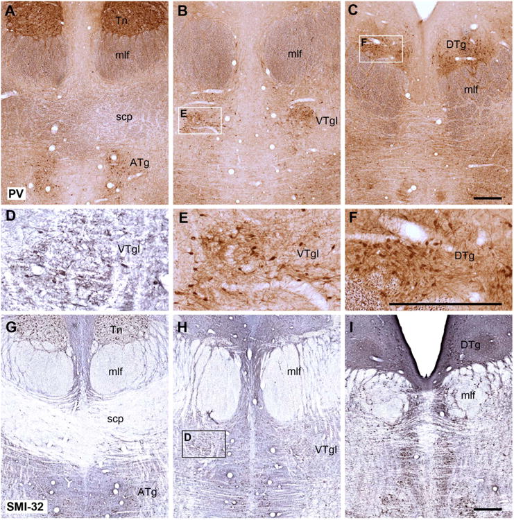
Coronal sections from a rhesus monkey brain showing the appearance of the dorsal and ventral tegmentum at three different anterior-posterior levels stained immunohistochemically for parvalbumin (A–C,E,F) and for SMI32 (D,G–I). The parvalbumin staining is of particular interest as it appears to show an affinity for Gudden's tegmental nuclei but not the adjacent raphe nucleus. Insets outlined in B, C, and H are shown at a higher magnification, such that E and F show parvalbumin-positive cells taken from B and C, respectively, whereas D shows SMI32-positive cells in the ventral tegmental nucleus taken from H. For abbreviations, see list. Scale bar = 0.5 mm in C (applies to A–C) F (applies to D–F), and I (applies to G–I).