
Figure 4.
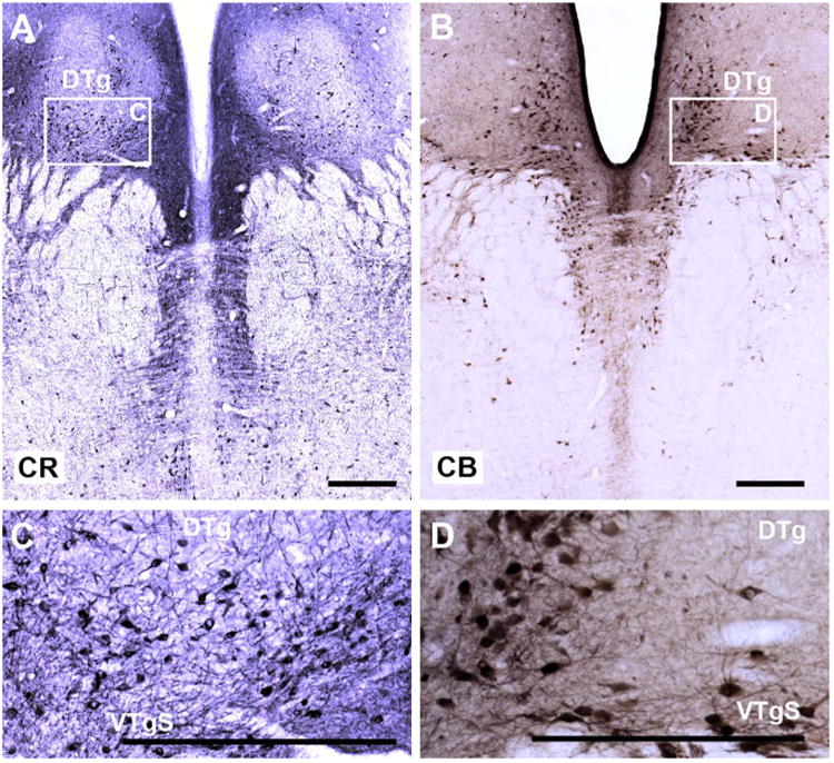
A–D: Coronal sections from a rhesus monkey brain showing the distribution of calretinin-positive cells (A,C) and calbindin-positive cells (B,D) in the tegmentum. As inset C shows, cells in both the dorsal tegmental and ventral tegmental nucleus are positive for calretinin, but this staining is not selective, as numerous raphe cells are also positive. Whereas cells in the ventral tegmentum also show a strong affinity for calbindin (B,D) this was not the case for the dorsal tegmentum. Again, adjacent cells in the dorsal raphe were immunopositive for calbindin. For abbreviations, see list. Scale bar = 0.5 mm in A–D.