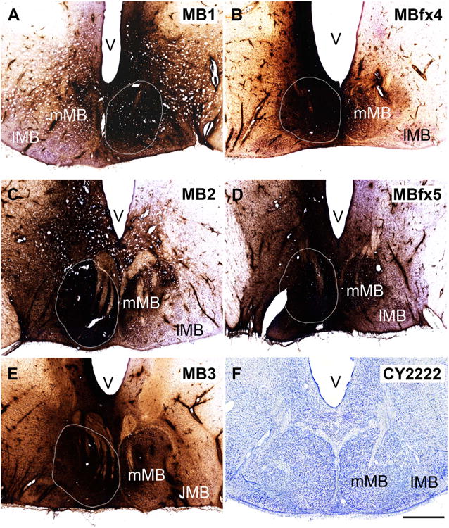
Figure 5.

A–F: Photomicrographs of the single injection sites within the mammillary bodies from the five cynomolgus macaques described in this study. The extent of the medial mammillary nucleus (mMB) is shown by a white outline. In all cases the injection was centered in the mMB, with variable spread into the lateral mammillary nucleus (lMB). The dark reaction product extends beyond the boundaries of the mammillary bodies, although the total extent of this product is appreciably greater than the active area of HRP uptake. F shows a coronal section through the lateral and medial mammillary bodies of a cynomolgus monkey (Nissl stain). V, ventricle. Scale bar = 1 mm in F (applies to a–F).
