
Figure 7.
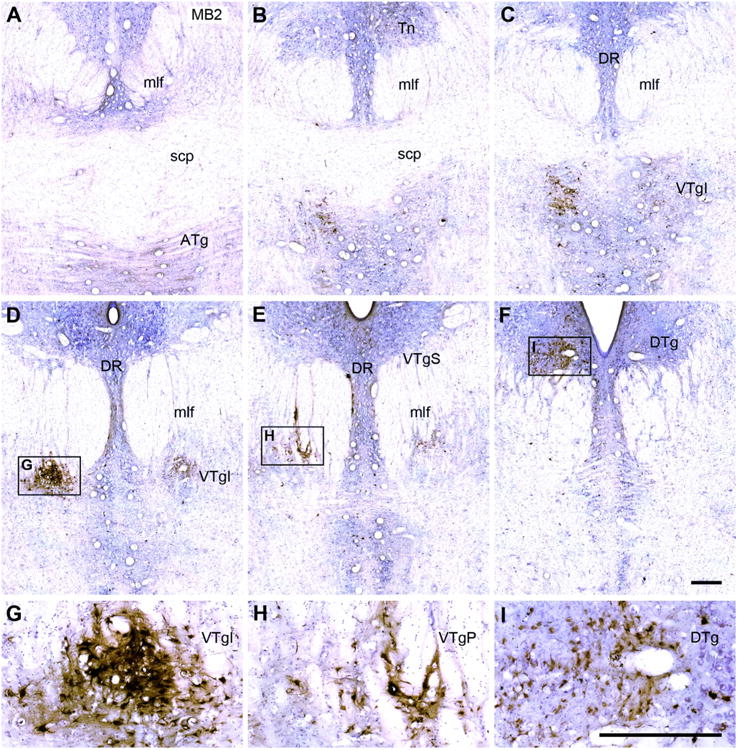
A–F: Six coronal Nissl-stained sections from case MB2 taken at different anterior (A) to posterior (F) levels through the tegmental area. The injection is presumed to have involved much of the medial and lateral mammillary nuclei. The HRP-positive cells are stained brown and form dense aggregates, especially in the ventral tegmental nucleus of Gudden. Note that the label “VTgI” in Figure 7C, D, and G is placed adjacent to the actual site of the nucleus. G,H,I: The HRP label has been magnified in these insets. Inset I shows HRP-positive cells primarily located in the dorsal tegmental nucleus of Gudden, pars centralis. For abbreviations, see list. Scale bar = 0.5 mm in I (applies to A–I).