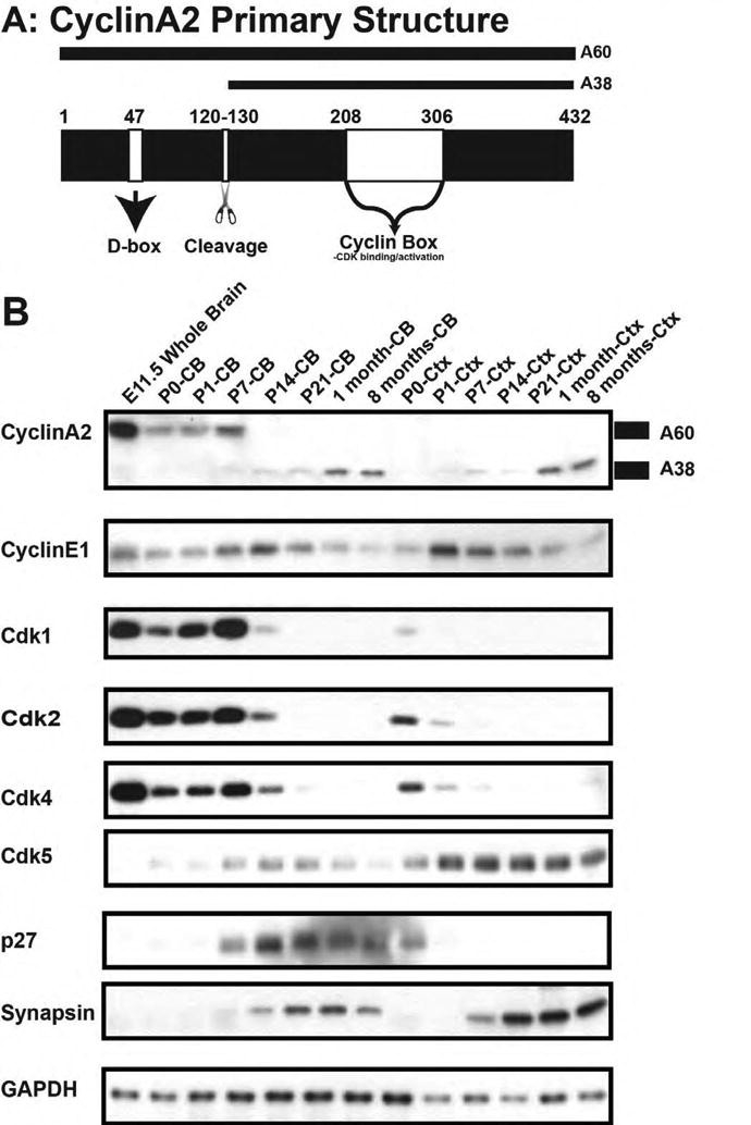
Figure 1. Cyclin A2 expression during CNS development.

(A) Schematic of cyclin A2 protein structure illustrated D-box, cleavage point, and CDK interacting domain. (B) Western blotting of forebrain and cerebellum during post-natal development. Cyclin A2 becomes processed into two forms (denoted on right of blot as A60 and A38). Top shows labels of post-natal dates, and on the left side of panel is the antibody used in the blot. Note that markers of proliferation cyclin A2, cdk1, cdk2, and cdk4 are robustly expressed in post-natal cerebellum whereas synapsin (a marker of differentiation) is seen at later post-natal ages (CB = cerebellum, CTX = cerebral cortex).
