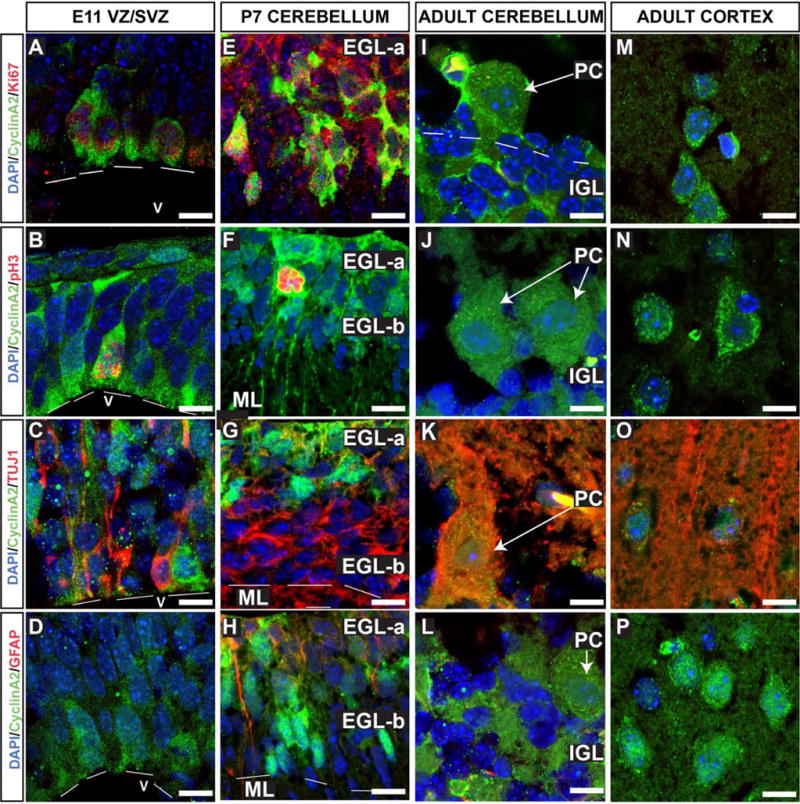
Figure 2. Cyclin A2 protein localization during DNS development.

Cryosections of fixed CD1 mouse embryos were stained for cyclin A2 in cells undergoing cell cycle (Ki67), mitotic cells (pH3), neurons (TUJ1), and astrocytes (GFAP). Sections were counterstained with DAPI to visualize nuclei (Blue). Animal ages and anatomical locations are depicted in the top row, and molecular markers are color coded on the left hand side. Cyclin A2 expression is noted in proliferating, undifferentiated neural stem cells at E11.5 (A-D). In the P7 cerebellum (E-H), cyclin A2 localizes to proliferating and mitotic cerebellar granule neuron progenitor cells and is excluded from differentiated neurons and astrocytes (E-H). In adult CNS, cyclin A2 is expressed in neurons. In panel B2, cerebellar folia numbers are placed in quotations (eg., “X”). (Scale bars = 10 μm, EGL = external granule layer, PC = Purkinje neuron, IGL = internal granule layer, ML = molecular layer, V = ventricle).
