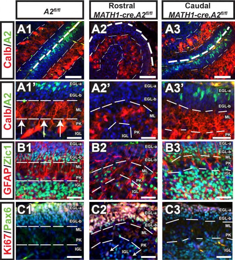
Figure 7. Intrinsic Proliferative Defects in CGNP's results in dyslamination of Purkinje neurons.

Genotypes and postnatal ages are denoted on the top, molecular markers are color coded and denoted on the left. Cyclin A2 was deleted in cerebellar granule precursor cells by intercrossing with Math1-cre mice and evaluated at P7 (genotypes are indicated atop each column). These mice showed incomplete cre-expression in caudal folia, which showed preserved cyclin A2 expression (A3). However, rostrally cyclin A2 was not detected in the EGL (A2). Calbindin staining in control shows laminar PC neurons, which in the cyclin A2 deleted areas showed PC neurons in dyslaminated aggregates (compare A1-A1′ to A2-A2′, and the internal control of A2, A3-A3′ ). Additional cytoarchitectural abnormalities included loss of Zic1+ cells in the EGL-b and a decrease in Zic1+ cells in the IGL (B1-B2). Pax6+Ki67+ cells were present in the full thickness of the EGL in the Math1-cre, cyclin A2fl/fl mice, which should normally show a band of Pax6+Ki67-negative cells in the EGL-b (C1-C2). White arrows in panel C3 point to Pax6+Ki67+ IGL cells. In the caudal folia of Math1-cre, A2fl/fl mice showed preserved cyclin A2 expression and showed cytoarchitecture similar to the control. Scale bars are as follows: A1-A3, B1-C3 = 50 μm, A1′-A3′ = 20 μm, The images show cerebella sectioned in the sagittal plane with the upper right being dorsal and the lower left being rostral (A2 = cyclin A2, Calb = calbindin).
